Odontogenic keratocyst: Difference between revisions
CSV import |
CSV import |
||
| Line 26: | Line 26: | ||
{{stub}} | {{stub}} | ||
<gallery> | |||
File:Keratocystic_odontogenic_tumour_-_2_-_very_high_mag.jpg|Odontogenic keratocyst under very high magnification | |||
File:Relative_incidence_of_odontogenic_cysts.jpg|Relative incidence of odontogenic cysts | |||
File:Classic_keratocystic_odontogenic_tumour.jpg|Classic keratocystic odontogenic tumour | |||
File:Keratocystic_odontogenic_tumour_-_2_-_intermed_mag.jpg|Odontogenic keratocyst under intermediate magnification | |||
File:Keratocystic_odontogenic_tumour_-_intermed_mag.jpg|Odontogenic keratocyst under intermediate magnification | |||
File:Keratocystic_odontogenic_tumour_-_2_-_high_mag.jpg|Odontogenic keratocyst under high magnification | |||
File:Massive_keratocystic_odontogenic_tumour.jpg|Massive keratocystic odontogenic tumour | |||
</gallery> | |||
Revision as of 11:35, 18 February 2025
Odontogenic keratocyst (OKC), also known as keratocystic odontogenic tumor (KCOT), is a rare and benign but locally aggressive developmental cystic neoplasm that arises from the dental lamina. It is characterized by its unique histopathological appearance and has a high recurrence rate.
Etiology
The exact cause of OKC is unknown. However, it is believed to be associated with the PTCH1 gene mutation. This mutation is also associated with nevoid basal cell carcinoma syndrome (NBCCS), a condition that increases the risk of developing multiple OKCs.
Clinical Features
OKCs can occur at any age but are most commonly diagnosed in the second and third decades of life. They have a slight male predilection and are more common in the mandible than the maxilla. Patients with OKC often present with swelling, pain, and occasionally pus discharge. However, many OKCs are asymptomatic and are discovered incidentally during routine dental radiographs.
Diagnosis
The diagnosis of OKC is based on a combination of clinical, radiographic, and histopathological findings. Radiographically, OKCs often appear as unilocular or multilocular radiolucent lesions with well-defined borders. Histologically, they are characterized by a thin, uniform epithelial lining with a corrugated parakeratin surface and a prominent basal cell layer.
Treatment
The treatment of OKC is controversial due to its high recurrence rate. Treatment options include conservative methods such as enucleation and curettage, and more aggressive methods such as resection. Recent studies have suggested the use of adjunctive therapies such as Carnoy's solution and cryotherapy to reduce the recurrence rate.
Prognosis
The prognosis of OKC is generally good with appropriate treatment. However, the high recurrence rate and association with NBCCS warrant long-term follow-up.
 | |
|---|---|
|
|
 |
-
 Odontogenic keratocyst under very high magnification
 -
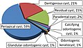 Relative incidence of odontogenic cysts
 -
 Classic keratocystic odontogenic tumour
 -
 Odontogenic keratocyst under intermediate magnification
 -
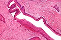 Odontogenic keratocyst under intermediate magnification
 -
 Odontogenic keratocyst under high magnification
 -
 Massive keratocystic odontogenic tumour


