Contrast CT: Difference between revisions
CSV import |
CSV import |
||
| Line 29: | Line 29: | ||
[[Category:Radiology]] | [[Category:Radiology]] | ||
{{medicine-stub}} | {{medicine-stub}} | ||
<gallery> | |||
File:Contrast_CT.jpg|Contrast CT | |||
File:SADDLE_PE.JPG|Saddle Pulmonary Embolism | |||
File:Volume_rendered.jpg|Volume Rendered Image | |||
File:CT_scan_of_hepatocellular_carcinoma,_without_and_with_IV_contrast.jpg|CT scan of hepatocellular carcinoma, without and with IV contrast | |||
</gallery> | |||
Latest revision as of 05:04, 18 February 2025
Contrast-enhanced computed tomography (contrast CT), also known as contrast-enhanced CT scan or simply contrast CT, is a medical imaging technique used in radiology to obtain detailed images of internal organs, blood vessels, and other tissues in the body. This method involves the use of computed tomography (CT) technology along with an intravenously injected contrast agent that enhances the visibility of certain structures or fluids within the body on the CT images.
Overview[edit]
Contrast CT is a pivotal diagnostic tool in medicine, offering enhanced visualization of bodily structures by utilizing contrast agents. These agents, typically iodine-based or barium-sulfate compounds, increase the X-ray absorption in the areas where they accumulate, making these regions appear different from their surroundings on the CT images. This difference in appearance helps in distinguishing between various types of tissues and in identifying abnormalities such as tumors, infections, and blood clots.
Procedure[edit]
The procedure for a contrast CT scan involves several steps. Initially, the patient may be asked to fast for a few hours before the scan. Upon arrival at the imaging center, an intravenous (IV) line is usually inserted into a vein in the patient's arm, through which the contrast material is injected. The rate and timing of the injection are carefully controlled to optimize the distribution of the contrast agent in the body.
Once the contrast material is administered, the patient is positioned on the CT scanner table, and the scan is performed. The CT scanner is a large machine with a tunnel in the center. The patient lies on a motorized table that slides into and out of this tunnel as the scanner rotates around the patient, capturing multiple cross-sectional images of the body.
Applications[edit]
Contrast CT scans are widely used in the diagnosis and management of various medical conditions. They are particularly useful in identifying and assessing:
- Cancers and tumors, by highlighting the blood vessels that feed the tumor
- Vascular diseases, including aneurysms, blockages, and atherosclerosis
- Infections, by showing areas of inflammation
- Trauma to organs and blood vessels
- Liver and kidney diseases
Risks and Considerations[edit]
While contrast CT scans are generally safe, there are some risks and considerations associated with their use. These include:
- Allergic reactions to the contrast material, ranging from mild (such as itching and hives) to severe (such as anaphylactic shock)
- Kidney damage, particularly in patients with pre-existing kidney problems or those with diabetes
- Radiation exposure, although modern CT scanners use the lowest possible dose of radiation that still provides high-quality images
Conclusion[edit]
Contrast-enhanced computed tomography is a valuable diagnostic tool in modern medicine, offering detailed and enhanced images of the body's internal structures. Despite its benefits, the use of contrast CT scans must be carefully considered by healthcare providers, taking into account the patient's health history and the potential risks associated with contrast agents and radiation exposure.
-
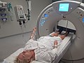 Contrast CT
 -
 Saddle Pulmonary Embolism
 -
Volume Rendered Image
-
CT scan of hepatocellular carcinoma, without and with IV contrast


