Brain herniation: Difference between revisions
mNo edit summary |
CSV import |
||
| Line 52: | Line 52: | ||
{{Medicine-stub}} | {{Medicine-stub}} | ||
<gallery> | |||
File:Decorticate.PNG|Decorticate posturing | |||
File:Brain_herniation_types-2.svg|Types of brain herniation | |||
File:Subfalcine-herniation-001.jpg|Subfalcine herniation | |||
File:Brain_injury_with_herniation_MRI.jpg|Brain injury with herniation on MRI | |||
</gallery> | |||
Revision as of 04:26, 18 February 2025
Brain herniation is a potentially fatal condition that occurs when parts of the brain are displaced from their usual position due to increased intracranial pressure. This displacement can compress brain structures and blood vessels, leading to decreased blood flow, oxygen deprivation, and further increase in intracranial pressure, creating a vicious cycle. Brain herniation can result from various causes, including traumatic brain injury, stroke, tumors, and infections.
Types
There are several types of brain herniation, each with distinct characteristics and implications for treatment and prognosis:
- Uncal herniation: This occurs when the uncus of the temporal lobe shifts downward, potentially compressing the brainstem and the third cranial nerve.
- Central herniation: In this type, the diencephalon and parts of the temporal lobes shift downward through the tentorial notch.
- Cingulate herniation: This involves the displacement of the cingulate gyrus under the falx cerebri.
- Transcalvarial herniation: Brain tissue herniates through a defect in the skull, often due to surgery or trauma.
- Tonsillar herniation: Also known as cerebellar or foramen magnum herniation, this occurs when the cerebellar tonsils move downward through the foramen magnum, potentially compressing the brainstem and leading to death.
Symptoms
Symptoms of brain herniation can vary depending on the type but may include:
- Changes in consciousness, ranging from drowsiness to coma
- Pupil dilation, particularly on one side, if the third cranial nerve is affected
- Changes in breathing patterns
- Weakness or paralysis on one side of the body
- Headache
- Seizures
Diagnosis
Diagnosis of brain herniation involves clinical assessment and imaging studies. Computed tomography (CT) scans and magnetic resonance imaging (MRI) are crucial for identifying the type and extent of herniation.
Treatment
Treatment of brain herniation is a medical emergency and focuses on reducing intracranial pressure, treating the underlying cause, and supporting vital functions. Interventions may include:
- Medications to reduce brain swelling, such as mannitol or hypertonic saline
- Ventriculostomy to drain cerebrospinal fluid and relieve pressure
- Surgical decompression to remove the source of increased pressure or to create more space for the swollen brain
Prognosis
The prognosis for brain herniation depends on the speed of diagnosis and treatment, the underlying cause, and the extent of brain damage. Early intervention can improve outcomes, but the condition can be fatal without prompt treatment.
Prevention
Preventing brain herniation involves managing conditions that can lead to increased intracranial pressure, such as head injuries, strokes, and brain tumors, with appropriate medical and surgical interventions.

-
 Decorticate posturing
 -
 Types of brain herniation
 -
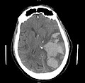 Subfalcine herniation
 -
 Brain injury with herniation on MRI


