Otic ganglion: Difference between revisions
CSV import |
CSV import |
||
| Line 31: | Line 31: | ||
{{Anatomy-stub}} | {{Anatomy-stub}} | ||
<gallery> | |||
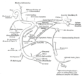
File:Gray782_updated.png|Otic ganglion and its connections | |||
File:Gray783.png|Otic ganglion, mandibular nerve, and branches | |||
File:Gray788.png|Otic ganglion and associated nerves | |||
File:Gray839.png|Otic ganglion and its anatomical relations | |||
</gallery> | |||
Latest revision as of 04:21, 18 February 2025
Otic Ganglion
The Otic ganglion is a small parasympathetic ganglion located immediately below the foramen ovale in the infratemporal fossa and on the medial surface of the mandibular nerve. It is functionally associated with the glossopharyngeal nerve and innervates the parotid gland for salivation.
Anatomy[edit]
The Otic ganglion is one of four parasympathetic ganglia of the head and neck. It is the smallest of the four and is located in the infratemporal fossa, immediately below the foramen ovale. The ganglion is positioned directly on the medial surface of the mandibular nerve (the V3 branch of the trigeminal nerve) and is connected to it by two small branches.
Function[edit]
The otic ganglion is functionally associated with the glossopharyngeal nerve. It receives preganglionic parasympathetic fibers from the glossopharyngeal nerve via the lesser petrosal nerve. These fibers synapse in the ganglion with postganglionic fibers that pass through the auriculotemporal nerve to reach the parotid gland, where they stimulate salivation.
Clinical significance[edit]
Damage to the otic ganglion can result in a loss of parasympathetic supply to the parotid gland. This can lead to a decrease in salivation, resulting in a dry mouth condition known as xerostomia.
See also[edit]
References[edit]
<references />

-
 Otic ganglion and its connections
 -
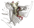 Otic ganglion, mandibular nerve, and branches
 -
 Otic ganglion and associated nerves
 -
 Otic ganglion and its anatomical relations


