Truncus arteriosus: Difference between revisions
CSV import |
CSV import |
||
| Line 43: | Line 43: | ||
[[Category:Congenital heart defects]] | [[Category:Congenital heart defects]] | ||
<gallery> | |||
File:Gray462.png|Diagram of the heart showing truncus arteriosus | |||
File:Gray469.png|Illustration of the heart with truncus arteriosus | |||
File:Gray1088.png|Heart anatomy with truncus arteriosus | |||
File:Truncus_arteriosus.jpg|Truncus arteriosus | |||
</gallery> | |||
Latest revision as of 04:20, 18 February 2025
A rare congenital heart defect
Truncus arteriosus is a rare type of congenital heart defect in which a single blood vessel (the truncus arteriosus) comes out of the right and left ventricles, instead of the normal two vessels (the pulmonary artery and the aorta). This condition results in a mixture of oxygenated and deoxygenated blood being circulated throughout the body.
Anatomy and Pathophysiology[edit]
In a normal heart, the pulmonary artery and the aorta are separate, with the pulmonary artery carrying deoxygenated blood from the right ventricle to the lungs, and the aorta carrying oxygenated blood from the left ventricle to the rest of the body. In truncus arteriosus, these two vessels are combined into a single vessel, which then branches into the pulmonary arteries and the aorta.
The condition is often associated with a ventricular septal defect (VSD), which is a hole between the right and left ventricles. This allows blood to mix between the two sides of the heart, leading to reduced oxygen levels in the blood that is pumped to the body.
Types[edit]
Truncus arteriosus is classified into several types based on the anatomy of the pulmonary arteries and their origin from the truncus. The most commonly used classification is the Collett and Edwards classification, which includes:
- Type I: A single pulmonary trunk arises from the truncus arteriosus and then divides into the left and right pulmonary arteries.
- Type II: The left and right pulmonary arteries arise separately but close to each other from the posterior aspect of the truncus.
- Type III: The left and right pulmonary arteries arise separately from the lateral aspects of the truncus.
Diagnosis[edit]
Diagnosis of truncus arteriosus is typically made using echocardiography, which can visualize the single arterial trunk and associated VSD. Other imaging techniques, such as cardiac MRI or CT scan, may be used to provide additional anatomical details.
Treatment[edit]
The treatment for truncus arteriosus is surgical. The goal of surgery is to separate the pulmonary and systemic circulations. This is usually done by closing the VSD and creating a conduit from the right ventricle to the pulmonary arteries. Early surgical intervention is critical to prevent complications such as pulmonary hypertension.
Prognosis[edit]
With advances in surgical techniques, the prognosis for individuals with truncus arteriosus has improved significantly. However, long-term follow-up is necessary to monitor for potential complications, such as conduit obstruction or valve dysfunction.
Related pages[edit]
References[edit]
- Hoffman, J.I.E., & Kaplan, S. (2002). The incidence of congenital heart disease. Journal of the American College of Cardiology, 39(12), 1890-1900.
- Van Praagh, R. (2009). The segmental approach to diagnosis in congenital heart disease. Pediatric Cardiology, 30(5), 701-705.
-
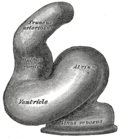 Diagram of the heart showing truncus arteriosus
 -
Illustration of the heart with truncus arteriosus
-
Heart anatomy with truncus arteriosus
-
Truncus arteriosus

