Tillaux fracture: Difference between revisions
CSV import Tags: mobile edit mobile web edit |
CSV import |
||
| Line 30: | Line 30: | ||
[[Category:Injuries]] | [[Category:Injuries]] | ||
{{medicine-stub}} | {{medicine-stub}} | ||
== Tillaux fracture == | |||
<gallery> | |||
File:Tillaux-Fraktur_11jw_-_Roe_ap_und_seitlich_-_001.png|X-ray images showing Tillaux fracture in AP and lateral views | |||
File:Tillaux-Fraktur_11jw_-_CT_cor_sag_ax_-_001.png|CT images showing Tillaux fracture in coronal, sagittal, and axial views | |||
</gallery> | |||
Revision as of 02:10, 18 February 2025
Tillaux Fracture
A Tillaux fracture is a specific type of ankle fracture that affects the anterolateral portion of the distal tibial epiphysis. It occurs due to an avulsion injury of the tibia at the attachment of the anterior tibiofibular ligament. This type of fracture is most commonly seen in adolescents and young adults, particularly those who engage in sports or high-impact activities. The injury mechanism typically involves a combination of rotation and axial load on the ankle, often when the foot is planted and the body twists.
Etiology and Pathophysiology
The Tillaux fracture is named after Paul Jules Tillaux, a French surgeon who first described this injury in 1892. The fracture occurs during a narrow window of skeletal development, usually between the ages of 12 and 15 years, when the medial part of the tibial epiphysis has fused, but the lateral part remains cartilaginous and susceptible to avulsion injuries. This vulnerability is due to the asymmetric closure of the growth plate, known as the physis, in the distal tibia.
Clinical Presentation
Patients with a Tillaux fracture typically present with pain, swelling, and limited mobility of the ankle joint following an injury. Physical examination may reveal tenderness over the anterolateral aspect of the ankle. It is crucial to differentiate this fracture from other types of ankle injuries, such as sprains or other fracture types, through detailed history and physical examination.
Diagnosis
Imaging studies are essential for diagnosing Tillaux fractures. Plain radiographs of the ankle, including anteroposterior, lateral, and mortise views, are usually the first step. These may show a fracture line extending from the tibial plafond into the distal tibial epiphysis. In some cases, computed tomography (CT) scans are required to fully assess the fracture's extent and displacement, which is crucial for planning treatment.
Treatment
The treatment of Tillaux fractures depends on the degree of displacement. Non-displaced or minimally displaced fractures can often be managed conservatively with immobilization in a cast or boot for a period of 4 to 6 weeks. However, displaced fractures require surgical intervention to realign the bone fragments and stabilize the ankle joint. Surgical techniques may include the use of screws or pins for fixation.
Prognosis
With appropriate treatment, the prognosis for a Tillaux fracture is generally good. Most patients can expect to return to their normal activities within 3 to 6 months. However, complications such as post-traumatic arthritis, growth disturbances, or instability of the ankle joint may occur, particularly if the fracture is not adequately reduced and stabilized.
Prevention
Preventing Tillaux fractures involves minimizing the risks associated with high-impact sports and activities. This includes proper training, use of protective gear, and adherence to safety guidelines. Early recognition and treatment of ankle injuries can also prevent more severe outcomes.

Tillaux fracture
-
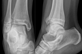 X-ray images showing Tillaux fracture in AP and lateral views
 -
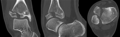 CT images showing Tillaux fracture in coronal, sagittal, and axial views

