Carotid sheath: Difference between revisions
CSV import Tags: mobile edit mobile web edit |
CSV import |
||
| Line 30: | Line 30: | ||
{{Anatomy-stub}} | {{Anatomy-stub}} | ||
<gallery> | |||
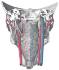
File:Carotid_sheath.PNG|Carotid sheath | |||
File:Gray794.png|Diagram of the carotid sheath and its contents | |||
File:Gray1031.png|Anatomy of the neck showing the carotid sheath | |||
</gallery> | |||
Latest revision as of 01:46, 18 February 2025
Carotid sheath is a tubular fascial layer that encloses several important structures within the neck. It is a key anatomical feature, providing a protective passage for the vascular and nervous components it contains. The carotid sheath is located on each side of the neck and extends from the base of the skull to the first rib and sternum.
Anatomy[edit]
The carotid sheath is composed of deep cervical fascia that envelops several vital structures:
- The common carotid artery and its branches, the internal carotid artery and external carotid artery
- The internal jugular vein
- The vagus nerve (Cranial Nerve X)
Additionally, the carotid sheath is in close relation to other significant anatomical structures, including the sympathetic trunk, which lies posteriorly to the sheath, and the ansa cervicalis, a nerve loop that is superficial to the sheath.
Function[edit]
The primary function of the carotid sheath is to provide a protective conduit for the structures it encases. This is crucial for the protection of major blood vessels and nerves that supply the head and neck. The sheath also serves to maintain the anatomical position of these structures, facilitating their proper functioning.
Clinical Significance[edit]
The carotid sheath is of significant interest in clinical medicine for several reasons:
- Carotid artery diseases: Conditions such as carotid artery stenosis or carotid artery dissection occur within the sheath and can lead to critical outcomes like stroke.
- Infections: Deep neck infections can spread within the carotid sheath, leading to severe complications.
- Surgical access: The carotid sheath is a landmark for surgeons during procedures like carotid endarterectomy or neck dissections for cancer.
Imaging[edit]
Imaging techniques such as ultrasound, computed tomography (CT) scans, and magnetic resonance imaging (MRI) are used to visualize the carotid sheath and its contents. These modalities are crucial for diagnosing diseases affecting the carotid sheath and for planning surgical interventions.
See Also[edit]

-
 Carotid sheath
 -
 Diagram of the carotid sheath and its contents
 -
 Anatomy of the neck showing the carotid sheath


