Clavipectoral triangle: Difference between revisions
CSV import Tags: mobile edit mobile web edit |
CSV import |
||
| Line 35: | Line 35: | ||
[[Category:Triangles]] | [[Category:Triangles]] | ||
[[Category:Medical terminology]] | [[Category:Medical terminology]] | ||
<gallery> | |||
File:Gray410.png | |||
File:Gray574.png | |||
</gallery> | |||
Latest revision as of 02:04, 17 February 2025
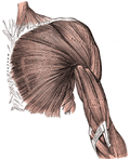
Clavipectoral Triangle (also known as Moynihan's triangle or subclavian triangle) is a small anatomical region of the upper chest. It is one of the four infraclavicular (below the clavicle) triangles.
Etymology[edit]
The term "clavipectoral" is derived from the Latin words "clavis" meaning key and "pectus" meaning chest. This is in reference to the triangle's location below the clavicle (key) and above the pectoralis major muscle (chest). The term "Moynihan's triangle" is named after the British surgeon, Berkeley Moynihan, who first described it.
Anatomy[edit]
The clavipectoral triangle is bounded by:
- Superiorly: The inferior border of the clavicle
- Medially: The lateral border of the sternocleidomastoid muscle
- Laterally: The medial border of the deltoid muscle
The floor of the triangle is formed by the pectoralis major muscle and the subclavius muscle. The roof is formed by the platysma muscle and the deep cervical fascia.
Contents[edit]
The clavipectoral triangle contains the following structures:
- The cephalic vein
- The thoracoacromial artery
- The lateral pectoral nerve
- Lymph nodes and fat
Clinical Significance[edit]
The clavipectoral triangle is an important landmark in surgical procedures involving the upper chest and shoulder. It is also used as a reference point in the diagnosis of certain medical conditions.
See Also[edit]
References[edit]
<references />


