Superior ophthalmic vein: Difference between revisions
CSV import |
CSV import |
||
| Line 29: | Line 29: | ||
{{stub}} | {{stub}} | ||
{{dictionary-stub1}} | {{dictionary-stub1}} | ||
== Superior ophthalmic vein == | |||
<gallery> | |||
File:Superior_ophthalmic_vein.png|Superior ophthalmic vein | |||
File:Gray572.png|Gray's Anatomy illustration 572 | |||
File:Gray570.png|Gray's Anatomy illustration 570 | |||
</gallery> | |||
Revision as of 01:56, 17 February 2025
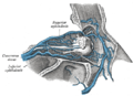
Superior ophthalmic vein is a vein that is located in the orbit of the eye. It is responsible for draining the frontal lobe and ethmoidal sinus, as well as the upper and medial parts of the retina. The superior ophthalmic vein is a significant part of the ophthalmic system and plays a crucial role in the overall health and function of the eye.
Anatomy
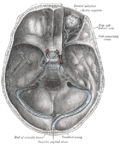
The superior ophthalmic vein originates in the medial angle of the orbit, a cavity in the skull that houses the eye. It is formed by the union of the veins of the nose and the supraorbital vein, which drains the forehead and scalp. The superior ophthalmic vein then passes through the superior orbital fissure, a gap in the skull, to drain into the cavernous sinus, a large vein at the base of the skull.
Function
The primary function of the superior ophthalmic vein is to drain blood from the upper and medial parts of the retina, the frontal lobe, and the ethmoidal sinus. This process is crucial for maintaining the health and function of the eye and surrounding structures.
Clinical significance
In some cases, the superior ophthalmic vein can become enlarged or dilated, a condition known as superior ophthalmic vein thrombosis. This can lead to symptoms such as proptosis (bulging of the eye), chemosis (swelling of the conjunctiva), and ophthalmoplegia (paralysis or weakness of the eye muscles). Treatment for this condition typically involves addressing the underlying cause, which can include infection, trauma, or tumor.
See also
References
<references />
 | |
|---|---|
|
|
 |
This medical article is a stub. You can help Wikipedia by adding missing information. |
Superior ophthalmic vein
-
Superior ophthalmic vein
-
 Gray's Anatomy illustration 572
 -
 Gray's Anatomy illustration 570


