Brenner tumour: Difference between revisions
CSV import |
CSV import Tags: mobile edit mobile web edit |
||
| Line 41: | Line 41: | ||
<gallery> | <gallery> | ||
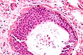
File:Brenner_tumour_intermed_mag.jpg|Brenner tumour | File:Brenner_tumour_intermed_mag.jpg|Brenner tumour | ||
File:Brenner_tumor_with_annotated_coffee_bean_nuclei.jpg|Brenner tumor with annotated coffee bean nuclei | |||
File:Walthard_cell_rest_-_very_high_mag.jpg|Walthard cell rest - very high magnification | |||
</gallery> | |||
<gallery> | |||
File:Brenner_Tumor_of_Ovary.jpg|Brenner tumor of ovary | |||
File:Brenner_tumour_intermed_mag.jpg|Brenner tumour intermediate magnification | |||
File:Brenner_tumor_with_annotated_coffee_bean_nuclei.jpg|Brenner tumor with annotated coffee bean nuclei | File:Brenner_tumor_with_annotated_coffee_bean_nuclei.jpg|Brenner tumor with annotated coffee bean nuclei | ||
File:Walthard_cell_rest_-_very_high_mag.jpg|Walthard cell rest - very high magnification | File:Walthard_cell_rest_-_very_high_mag.jpg|Walthard cell rest - very high magnification | ||
</gallery> | </gallery> | ||
Revision as of 04:57, 18 February 2025
Brenner tumour is a rare type of ovarian neoplasm that is part of the surface epithelial-stromal tumour group of ovarian cancers. It was first described by Robert Brenner in 1907.
Overview
Brenner tumours are typically benign, but can sometimes be malignant or borderline. They are usually asymptomatic and are often discovered incidentally during routine gynaecological examinations.
Pathology
Brenner tumours are composed of small, round, uniform cells that resemble transitional epithelium (the type of tissue that lines the urinary tract). These cells are typically arranged in nests surrounded by dense fibrous tissue.
Clinical Features
Most Brenner tumours are asymptomatic, but some women may experience pelvic pain or pressure. On physical examination, a pelvic mass may be palpable.
Diagnosis
The diagnosis of Brenner tumour is usually made based on the histological appearance of the tumour. Imaging studies such as ultrasound, CT scan, or MRI may also be used to identify the tumour.
Treatment
The treatment for Brenner tumour is usually surgical removal of the tumour. In some cases, chemotherapy or radiation therapy may be used if the tumour is malignant.
Prognosis
The prognosis for women with Brenner tumour is generally good, especially if the tumour is benign. However, malignant Brenner tumours can be more aggressive and have a poorer prognosis.
See Also
References
<references />
 | |
|---|---|
|
|
 |
-
 Brenner tumour
 -
 Brenner tumor with annotated coffee bean nuclei
 -
 Walthard cell rest - very high magnification


-
 Brenner tumor of ovary
 -
Brenner tumour intermediate magnification
-
Brenner tumor with annotated coffee bean nuclei
-
Walthard cell rest - very high magnification

