Transthoracic echocardiogram: Difference between revisions
CSV import |
CSV import |
||
| Line 66: | Line 66: | ||
[[Category:Cardiology]] | [[Category:Cardiology]] | ||
[[Category:Medical imaging]] | [[Category:Medical imaging]] | ||
<gallery> | |||
File:Echocardiogram_in_US_Navy.jpg|Transthoracic echocardiogram | |||
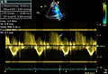
File:Doppler_mitral_valve.gif|Transthoracic echocardiogram | |||
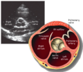
File:Flow_in_pulmonic_valve.jpg|Transthoracic echocardiogram | |||
File:LeftParasternalLongAxis.gif|Transthoracic echocardiogram | |||
File:LeftVentricleShortAxis.gif|Transthoracic echocardiogram | |||
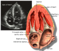
File:Aortic_valve_sa.png|Transthoracic echocardiogram | |||
File:Apical_4_chamber_view.png|Transthoracic echocardiogram | |||
File:Apical2Chamber.png|Transthoracic echocardiogram | |||
File:Subcostal_view_of_heart.gif|Transthoracic echocardiogram | |||
File:Fig009_(CardioNetworks_ECHOpedia).svg|Transthoracic echocardiogram | |||
</gallery> | |||
Latest revision as of 12:16, 18 February 2025
Transthoracic Echocardiogram[edit]
A transthoracic echocardiogram (TTE) is a non-invasive medical test that uses ultrasound waves to create images of the heart. It is one of the most common types of echocardiography and is used to assess the heart's structure and function.
Procedure[edit]
During a transthoracic echocardiogram, a sonographer or a cardiologist applies a gel to the patient's chest and uses a transducer to send ultrasound waves through the chest wall. These waves bounce off the heart structures and are captured by the transducer, which sends the data to a computer to create images of the heart.
Uses[edit]
Transthoracic echocardiograms are used to evaluate:
- The size and shape of the heart chambers
- The movement of the heart walls
- The function of the heart valves
- The presence of any abnormal masses or fluid around the heart
Images[edit]


.svg)
Advantages[edit]
The transthoracic echocardiogram is a widely used diagnostic tool because it is:
- Non-invasive
- Safe and painless
- Provides real-time images
- Can be performed at the bedside
Limitations[edit]
While TTE is a valuable diagnostic tool, it has some limitations, including:
- Limited image quality in patients with obesity or lung disease
- Difficulty in visualizing certain heart structures
Related Pages[edit]
References[edit]
-
 Transthoracic echocardiogram
 -
 Transthoracic echocardiogram
 -
 Transthoracic echocardiogram
 -
 Transthoracic echocardiogram
 -
 Transthoracic echocardiogram
 -
 Transthoracic echocardiogram
 -
 Transthoracic echocardiogram
 -
 Transthoracic echocardiogram
 -
 Transthoracic echocardiogram
 -
 Transthoracic echocardiogram
