Glandular odontogenic cyst: Difference between revisions
CSV import |
CSV import |
||
| Line 62: | Line 62: | ||
[[Category:Oral pathology]] | [[Category:Oral pathology]] | ||
[[Category:Rare diseases]] | [[Category:Rare diseases]] | ||
== Glandular_odontogenic_cyst == | |||
<gallery> | |||
File:Relative_incidence_of_odontogenic_cysts.jpg|Relative incidence of odontogenic cysts | |||
File:PDB_1ysw_EBI.jpg|PDB 1ysw EBI | |||
File:Panoramic_Xray.jpg|Panoramic X-ray | |||
</gallery> | |||
Latest revision as of 21:24, 23 February 2025
| Glandular odontogenic cyst | |
|---|---|
| Synonyms | |
| Pronounce | N/A |
| Specialty | N/A |
| Symptoms | Swelling, pain, displacement of teeth |
| Complications | Potential for aggressive growth |
| Onset | |
| Duration | |
| Types | |
| Causes | Unknown |
| Risks | |
| Diagnosis | Clinical examination, radiographic imaging, histopathological analysis |
| Differential diagnosis | Odontogenic keratocyst, Ameloblastoma, Dentigerous cyst |
| Prevention | N/A |
| Treatment | Surgical enucleation, curettage |
| Medication | |
| Prognosis | Generally good with treatment |
| Frequency | Rare |
| Deaths | N/A |
The glandular odontogenic cyst (GOC) is a rare type of odontogenic cyst that occurs in the jaws. It is characterized by its unique histological features and potential for aggressive behavior.
Epidemiology[edit]
Glandular odontogenic cysts are rare, accounting for approximately 0.2% of all odontogenic cysts. They can occur in a wide age range, but are most commonly diagnosed in adults between the ages of 40 and 60. There is a slight male predilection.
Clinical Presentation[edit]
Patients with a glandular odontogenic cyst may present with a painless swelling in the jaw, although some may experience pain or discomfort. The cyst can cause displacement of teeth and, in some cases, resorption of adjacent bone. It most commonly occurs in the anterior region of the mandible, but can also be found in the maxilla.
Pathogenesis[edit]
The exact cause of glandular odontogenic cysts is unknown. They are believed to arise from the odontogenic epithelium, which is the tissue involved in tooth development. The cysts are characterized by their glandular or salivary-like features, which are unique among odontogenic cysts.
Histopathology[edit]
Histologically, glandular odontogenic cysts exhibit a lining of non-keratinized stratified squamous epithelium with areas of columnar or cuboidal cells. The epithelium may show "hobnail" cells, mucous cells, and microcystic spaces. These features can resemble salivary gland tissue, hence the name "glandular" odontogenic cyst.
Radiographic Features[edit]
On radiographic examination, glandular odontogenic cysts typically appear as well-defined radiolucent lesions. They may be unilocular or multilocular, and can cause expansion of the cortical bone. The radiographic appearance can be similar to other odontogenic cysts and tumors, making histopathological examination essential for diagnosis.
Diagnosis[edit]
The diagnosis of a glandular odontogenic cyst is based on a combination of clinical, radiographic, and histopathological findings. A biopsy is necessary to confirm the diagnosis and differentiate it from other lesions such as the odontogenic keratocyst or ameloblastoma.
Differential Diagnosis[edit]
The differential diagnosis for glandular odontogenic cyst includes:
Treatment[edit]
The treatment of choice for glandular odontogenic cysts is surgical enucleation and curettage. Due to the potential for recurrence, careful follow-up is recommended. In some cases, more aggressive surgical approaches may be necessary.
Prognosis[edit]
The prognosis for patients with glandular odontogenic cysts is generally good following appropriate surgical treatment. However, there is a risk of recurrence, which necessitates long-term follow-up.
See Also[edit]
Template:Oral and maxillofacial pathology
Glandular_odontogenic_cyst[edit]
-
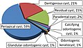 Relative incidence of odontogenic cysts
 -
 PDB 1ysw EBI
 -
 Panoramic X-ray


