Preclinical SPECT: Difference between revisions
CSV import |
CSV import |
||
| Line 21: | Line 21: | ||
{{medicine-stub}} | {{medicine-stub}} | ||
<gallery> | |||
File:High_resolution_mouse_bone_SPECT_scan.gif|High resolution mouse bone SPECT scan | |||
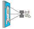
File:Pinhole_imaging.jpg|Pinhole imaging | |||
File:Mouse_SPECT_scan_-_circulation_of_radiolabeled_nanoparticles.gif|Mouse SPECT scan - circulation of radiolabeled nanoparticles | |||
File:15_secod_TB.gif|15 second TB | |||
File:Matrix_of_medical_imaging.jpg|Matrix of medical imaging | |||
</gallery> | |||
Latest revision as of 04:32, 18 February 2025
Preclinical Single Photon Emission Computed Tomography (Preclinical SPECT) is a nuclear medicine imaging technique that is used extensively in the field of preclinical research. Preclinical SPECT allows for the non-invasive visualization, quantification, and tracking of biological processes at the cellular and molecular level in living subjects, primarily in small animals such as rodents. This technology plays a crucial role in the development of new pharmaceuticals, the study of disease mechanisms, and the evaluation of new therapeutic strategies before they are tested in humans.
Overview[edit]
Preclinical SPECT works on the principle of detecting gamma rays emitted from radioactively labeled compounds, known as radiotracers, which are injected into the subject. These radiotracers are designed to target specific biological processes. The gamma rays are detected by a gamma camera, and the data collected is then reconstructed to create three-dimensional images that represent the distribution of the radiotracer within the body of the subject.
Components and Functioning[edit]
The core components of a Preclinical SPECT system include a gamma camera, a computer for image reconstruction, and software for data analysis. The gamma camera consists of one or more detectors that rotate around the subject, capturing gamma rays from multiple angles. Advanced systems may employ pinhole or multipinhole collimators to achieve higher spatial resolution, which is critical for imaging small animals.
Applications[edit]
Preclinical SPECT is utilized in various research areas including, but not limited to, oncology, neurology, cardiology, and pharmacology. In oncology, it can be used to monitor tumor growth, metastasis, and response to therapy. In neurology, it aids in understanding neurological disorders such as Alzheimer's disease by visualizing changes in brain metabolism and neurotransmitter systems. In cardiology, it helps in assessing myocardial perfusion and function. Furthermore, in pharmacology, Preclinical SPECT provides valuable information on the pharmacokinetics and pharmacodynamics of new drugs.
Advantages and Limitations[edit]
One of the main advantages of Preclinical SPECT is its ability to provide in vivo imaging with high sensitivity and specificity. It allows for longitudinal studies on the same subjects, reducing the number of animals required for research and providing more consistent data. However, its limitations include relatively lower spatial resolution compared to other imaging modalities such as micro-CT or micro-MRI, and the necessity of using radioactive materials, which requires special handling and safety measures.
Future Directions[edit]
The future of Preclinical SPECT includes the development of new radiotracers that can target a wider range of biological processes, improvements in detector technology to enhance image quality and resolution, and the integration with other imaging modalities such as CT or MRI to provide complementary anatomical and functional information. This multimodal imaging approach can significantly enhance the understanding of complex biological systems and diseases.

-
 High resolution mouse bone SPECT scan
 -
 Pinhole imaging
 -
 Mouse SPECT scan - circulation of radiolabeled nanoparticles
 -
15 second TB
-
Matrix of medical imaging

