Craniofacial cleft: Difference between revisions
CSV import Tags: mobile edit mobile web edit |
CSV import |
||
| Line 38: | Line 38: | ||
{{Medicine-stub}} | {{Medicine-stub}} | ||
<gallery> | |||
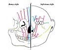
File:Picture Tessier classification.jpg|Craniofacial cleft | |||
File:Gould Pyle 101.jpg|Craniofacial cleft | |||
File:Gould Pyle 102.jpg|Craniofacial cleft | |||
File:Van der meulen - internasal dysplasia.jpg|Craniofacial cleft | |||
File:Nasal dysplasia.jpg|Craniofacial cleft | |||
File:Nasalmaxillary dysplasia.jpg|Craniofacial cleft | |||
File:Maxillary dysplasia.jpg|Craniofacial cleft | |||
File:Picture box osteotomy.jpg|Craniofacial cleft | |||
File:Picture median fasciotomy.jpg|Craniofacial cleft | |||
</gallery> | |||
Latest revision as of 01:22, 20 February 2025
Craniofacial cleft is a congenital deformity characterized by an abnormal gap or cleft in the face and/or the cranium. These clefts can vary greatly in size, shape, and location, affecting not only the appearance and function of the face but also potentially impacting the brain and the development of the skull. Craniofacial clefts are part of a broader category of craniofacial anomalies and can occur as isolated defects or as part of a syndrome.
Etiology[edit]
The exact cause of craniofacial clefts is not fully understood, but it is believed to be a combination of genetic and environmental factors. Disruptions in the normal development of the face and skull during embryonic growth lead to these anomalies. Factors that may increase the risk include genetic mutations, maternal smoking, use of certain medications during pregnancy, and nutritional deficiencies.
Classification[edit]
Craniofacial clefts are classified according to their location and the structures they involve. The most widely used classification system is the Tessier cleft classification system, which numbers clefts from 0 to 14, based on their anatomical position. This system helps in understanding the complexity of the clefts and planning for surgical intervention.
Symptoms and Complications[edit]
The symptoms of craniofacial clefts can vary widely depending on the severity and location of the cleft. Common issues include:
- Difficulty with feeding and swallowing
- Breathing problems
- Speech difficulties
- Hearing loss
- Dental problems
- Visual impairments
Complications may also arise from associated anomalies, such as heart defects, brain abnormalities, and limb deformities in syndromic cases.
Diagnosis[edit]
Diagnosis of craniofacial clefts typically occurs through prenatal imaging, such as ultrasound, and is confirmed after birth by physical examination. Advanced imaging techniques, like CT scans and MRI, are used to assess the extent of the cleft and plan for surgical correction.
Treatment[edit]
Treatment of craniofacial clefts is complex and often requires a multidisciplinary approach, involving surgeons, pediatricians, dentists, speech therapists, and other specialists. The primary goal of treatment is to improve function and appearance. This often involves multiple surgeries over the course of several years, including reconstructive surgery to close the cleft, orthodontic treatment, and speech therapy.
Prognosis[edit]
The prognosis for individuals with craniofacial clefts varies depending on the severity of the cleft and the presence of associated anomalies. With early and comprehensive treatment, many individuals can lead healthy, fulfilling lives.
See Also[edit]

-
 Craniofacial cleft
 -
 Craniofacial cleft
 -
 Craniofacial cleft
 -
 Craniofacial cleft
 -
 Craniofacial cleft
 -
 Craniofacial cleft
 -
 Craniofacial cleft
 -
 Craniofacial cleft
 -
 Craniofacial cleft


