Tissue microarray: Difference between revisions
CSV import Tags: mobile edit mobile web edit |
CSV import |
||
| Line 24: | Line 24: | ||
{{Medicine-stub}} | {{Medicine-stub}} | ||
<gallery> | |||
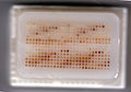
File:Tissue_MicroArray_Block.jpg|Tissue MicroArray Block | |||
File:0.6_mm_core_Tissue_MicroArray_Block.jpg|0.6 mm core Tissue MicroArray Block | |||
File:Tissue_MicroArray_Slide.jpg|Tissue MicroArray Slide | |||
</gallery> | |||
Latest revision as of 03:54, 18 February 2025
Tissue Microarray (TMA) is a high-throughput technology used in pathology and cancer research for the analysis of multiple tissue samples simultaneously. It involves the extraction of tissue cores from various donor blocks and their re-arrangement into a new, single recipient block. This technique allows for the rapid visualization and assessment of molecular markers within a large number of tissue specimens, facilitating comparative studies across different tissue types or disease states.
Overview[edit]
The concept of TMA was developed to address the need for a more efficient way of conducting studies on tissue specimens. Traditional methods, which involve the examination of one slide at a time, are time-consuming and resource-intensive. TMA technology, on the other hand, enables researchers to analyze hundreds of tissue samples on a single slide. This not only conserves valuable tissue but also standardizes experimental conditions, improving the reliability of comparative analyses.
Construction[edit]
The construction of a TMA involves several key steps. First, donor tissue blocks and corresponding histological slides are reviewed to identify representative areas for sampling. Using a tissue arrayer, cylindrical cores (typically 0.6mm to 2mm in diameter) are punched from these selected areas of the donor blocks and inserted into a pre-drilled recipient block in a precisely organized array. Once constructed, the TMA block can be sectioned into thin slices using a microtome, and these sections are then transferred to slides for subsequent analysis.
Applications[edit]
TMA technology has a wide range of applications, particularly in the field of oncology. It is used for the validation of novel biomarkers, the study of cancer epidemiology, and the assessment of gene expression patterns. TMAs are also valuable in drug discovery and development, as they can be used to evaluate the efficacy of therapeutic agents across different tissue types or disease states. Furthermore, TMAs facilitate the study of tissue-specific expression of proteins, helping to elucidate the molecular mechanisms underlying various diseases.
Advantages and Limitations[edit]
One of the main advantages of TMA is its efficiency. By allowing for the simultaneous analysis of multiple samples, it significantly reduces the time and resources required for large-scale studies. Additionally, the use of standardized sections ensures that experimental conditions are consistent across all samples, enhancing the comparability of results.
However, TMA technology also has its limitations. The small size of the tissue cores may not be representative of the entire donor tissue block, potentially leading to sampling bias. Moreover, the quality of the TMA sections can be affected by the quality of the donor tissue and the precision of the array construction process.
Conclusion[edit]
Tissue Microarray is a powerful tool in the field of pathology and cancer research, offering a high-throughput method for the analysis of tissue specimens. Despite its limitations, the technology has significantly contributed to the advancement of molecular pathology, providing valuable insights into the molecular basis of diseases and facilitating the development of targeted therapies.

-
 Tissue MicroArray Block
 -
 0.6 mm core Tissue MicroArray Block
 -
 Tissue MicroArray Slide


