Arenga pinnata: Difference between revisions
CSV import |
CSV import |
||
| Line 1: | Line 1: | ||
{{Short description|Congenital heart defect}} | |||
{{Use dmy dates|date=October 2023}} | |||
== | ==Aortopulmonary window== | ||
The ''' | The '''aortopulmonary window''' is a rare congenital heart defect characterized by an abnormal communication between the [[aorta]] and the [[pulmonary artery]]. This defect allows oxygen-rich blood from the aorta to mix with oxygen-poor blood in the pulmonary artery, leading to increased blood flow to the lungs and potentially causing pulmonary hypertension and heart failure if left untreated. | ||
== | ==Anatomy and Pathophysiology== | ||
The | The aortopulmonary window is a defect in the septum that normally separates the aorta and the pulmonary artery. This septum is part of the embryonic truncus arteriosus, which normally divides into the aorta and pulmonary artery during fetal development. Failure of this septum to form properly results in a communication between these two major vessels. | ||
The size of the defect can vary, and the clinical presentation depends on the size of the window and the amount of blood shunting from the aorta to the pulmonary artery. A large defect can lead to significant left-to-right shunting, causing volume overload of the pulmonary circulation and the left side of the heart. | |||
The | |||
== | ==Clinical Presentation== | ||
Patients with an aortopulmonary window may present with symptoms of heart failure, such as difficulty breathing, poor feeding, and failure to thrive in infants. Older children may experience exercise intolerance and frequent respiratory infections. On physical examination, a continuous murmur may be heard due to the abnormal blood flow between the aorta and pulmonary artery. | |||
== | ==Diagnosis== | ||
The diagnosis of an aortopulmonary window is typically made using [[echocardiography]], which can visualize the defect and assess the degree of shunting. Additional imaging studies, such as [[cardiac MRI]] or [[CT angiography]], may be used to provide further anatomical details. | |||
== | ==Treatment== | ||
Surgical repair is the definitive treatment for an aortopulmonary window. The procedure involves closing the defect with a patch, which separates the aorta and pulmonary artery and eliminates the abnormal shunt. Early surgical intervention is recommended to prevent the development of irreversible pulmonary vascular disease. | |||
==Prognosis== | |||
With timely surgical repair, the prognosis for patients with an aortopulmonary window is generally good. Most patients can expect normal growth and development following successful closure of the defect. However, if left untreated, the condition can lead to significant morbidity and mortality due to complications such as pulmonary hypertension and heart failure. | |||
[[ | ==Related pages== | ||
[[ | * [[Congenital heart defect]] | ||
[[Category: | * [[Patent ductus arteriosus]] | ||
* [[Ventricular septal defect]] | |||
* [[Pulmonary hypertension]] | |||
==Gallery== | |||
<gallery> | |||
File:Aortopulmonary_window.svg|Diagram of an aortopulmonary window | |||
</gallery> | |||
[[Category:Congenital heart defects]] | |||
Revision as of 19:23, 11 February 2025
Congenital heart defect
Aortopulmonary window
The aortopulmonary window is a rare congenital heart defect characterized by an abnormal communication between the aorta and the pulmonary artery. This defect allows oxygen-rich blood from the aorta to mix with oxygen-poor blood in the pulmonary artery, leading to increased blood flow to the lungs and potentially causing pulmonary hypertension and heart failure if left untreated.
Anatomy and Pathophysiology
The aortopulmonary window is a defect in the septum that normally separates the aorta and the pulmonary artery. This septum is part of the embryonic truncus arteriosus, which normally divides into the aorta and pulmonary artery during fetal development. Failure of this septum to form properly results in a communication between these two major vessels.
The size of the defect can vary, and the clinical presentation depends on the size of the window and the amount of blood shunting from the aorta to the pulmonary artery. A large defect can lead to significant left-to-right shunting, causing volume overload of the pulmonary circulation and the left side of the heart.
Clinical Presentation
Patients with an aortopulmonary window may present with symptoms of heart failure, such as difficulty breathing, poor feeding, and failure to thrive in infants. Older children may experience exercise intolerance and frequent respiratory infections. On physical examination, a continuous murmur may be heard due to the abnormal blood flow between the aorta and pulmonary artery.
Diagnosis
The diagnosis of an aortopulmonary window is typically made using echocardiography, which can visualize the defect and assess the degree of shunting. Additional imaging studies, such as cardiac MRI or CT angiography, may be used to provide further anatomical details.
Treatment
Surgical repair is the definitive treatment for an aortopulmonary window. The procedure involves closing the defect with a patch, which separates the aorta and pulmonary artery and eliminates the abnormal shunt. Early surgical intervention is recommended to prevent the development of irreversible pulmonary vascular disease.
Prognosis
With timely surgical repair, the prognosis for patients with an aortopulmonary window is generally good. Most patients can expect normal growth and development following successful closure of the defect. However, if left untreated, the condition can lead to significant morbidity and mortality due to complications such as pulmonary hypertension and heart failure.
Related pages
Gallery
-
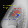 Diagram of an aortopulmonary window

