Zygomatic arch: Difference between revisions
CSV import |
CSV import |
||
| Line 32: | Line 32: | ||
{{stub}} | {{stub}} | ||
<gallery> | |||
File:Gray188-Sphenozygomatic_suture.png|Sphenozygomatic suture | |||
File:Processuszygomaticusossisfrontalis.PNG|Processus zygomaticus ossis frontalis | |||
File:Zygomatic-arch.jpg|Zygomatic arch | |||
File:Gray137.png|Zygomatic arch | |||
File:Gray165.png|Zygomatic arch | |||
File:Gray187.png|Zygomatic arch | |||
File:Gray382.png|Zygomatic arch | |||
File:Gray1024.png|Zygomatic arch | |||
File:Sygomafracture.png|Zygomatic arch fracture | |||
File:Slide6JAN.JPG|Zygomatic arch | |||
</gallery> | |||
Latest revision as of 11:24, 18 February 2025
Zygomatic Arch
The Zygomatic arch or cheekbone is a bony arch in the skull that extends along the side of the skull and forms part of the orbit (eye socket). It is formed by the zygomatic process of the temporal bone and the temporal process of the zygomatic bone, the two meeting at the zygomaticotemporal suture.
Structure[edit]
The zygomatic arch is formed by the union of two processes: the zygomatic process of the temporal bone and the temporal process of the zygomatic bone. These two processes join at the zygomaticotemporal suture. The arch is palpable from the exterior, and it creates the prominence of the cheek, or the malar eminence.
Function[edit]
The zygomatic arch provides an attachment for the masseter muscle, which is one of the muscles involved in mastication (chewing). It also forms part of the lateral wall and floor of the orbit.
Clinical significance[edit]
Injury to the zygomatic arch can result in fractures and can affect the function of the masseter muscle, potentially affecting the ability to chew. Such injuries may also affect the appearance of the cheek, as the arch creates the prominence of the cheek.
See also[edit]
References[edit]
<references />
External links[edit]
|
|
 |
-
 Sphenozygomatic suture
 -
 Processus zygomaticus ossis frontalis
 -
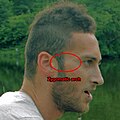 Zygomatic arch
 -
 Zygomatic arch
 -
 Zygomatic arch
 -
 Zygomatic arch
 -
 Zygomatic arch
 -
 Zygomatic arch
 -
 Zygomatic arch fracture
 -
Zygomatic arch


