Fluorescence microscope: Difference between revisions
CSV import |
CSV import |
||
| Line 40: | Line 40: | ||
{{stub}} | {{stub}} | ||
<gallery> | |||
File:Olympus-BX61-fluorescence_microscope.jpg|Olympus BX61 fluorescence microscope | |||
File:FluorescenceFilters_2008-09-28.svg|Fluorescence Filters 2008-09-28 | |||
File:FluorescenceMicroscopeSample_HerringSpermSYBRGreen.jpg|Fluorescence Microscope Sample Herring Sperm SYBR Green | |||
File:Depth_Coded_Phalloidin_Stained_Actin_Filaments_Cancer_Cell.png|Depth Coded Phalloidin Stained Actin Filaments Cancer Cell | |||
File:Dividing_Cell_Fluorescence.jpg|Dividing Cell Fluorescence | |||
File:FluorescentCells.jpg|Fluorescent Cells | |||
File:3D_Dual_Color_Super_Resolution_Microscopy_Cremer_2010.png|3D Dual Color Super Resolution Microscopy Cremer 2010 | |||
File:FISH_13_21.jpg|FISH 13 21 | |||
File:Yeast_membrane_proteins.jpg|Yeast membrane proteins | |||
</gallery> | |||
Latest revision as of 21:23, 23 February 2025
Fluorescence microscope is a specific type of microscope that uses fluorescence and phosphorescence instead of, or in addition to, reflection and absorption to study properties of organic or inorganic substances.
History[edit]
The fluorescence microscope was first developed by August Köhler in 1904. It was further improved by Oskar Heimstädt, who published the first paper on fluorescence microscopy in 1911.
Principle[edit]
The basic task of the fluorescence microscope is to let excitation light radiate the specimen and then sort out the much weaker emitted light to make up the image. The main components of the microscope are the light source, the excitation filter, the dichroic mirror, and the emission filter.
Types of Fluorescence Microscopes[edit]
There are several types of fluorescence microscopes, including:
- Epifluorescence microscope: This is the most common type of fluorescence microscope. It uses a dichroic mirror to direct the excitation light onto the specimen and then collect the fluorescence light.
- Confocal microscope: This microscope uses a laser to excite a single plane of the specimen and then uses a pinhole to eliminate out-of-focus light.
- Two-photon excitation microscope: This microscope uses two photons of lower energy to excite the specimen. This allows for deeper penetration into the specimen and reduces photobleaching.
- Super-resolution microscope: This microscope surpasses the diffraction limit of light to provide higher resolution images.
Applications[edit]
Fluorescence microscopes are used in various fields, including biology, medicine, and material science. They are particularly useful in studying cellular structures and processes, protein interactions, and understanding the internal structure of materials.
See Also[edit]
References[edit]
<references />
 | |
|---|---|
|
|
 |
-
 Olympus BX61 fluorescence microscope
 -
 Fluorescence Filters 2008-09-28
 -
 Fluorescence Microscope Sample Herring Sperm SYBR Green
 -
 Depth Coded Phalloidin Stained Actin Filaments Cancer Cell
 -
 Dividing Cell Fluorescence
 -
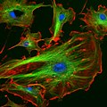 Fluorescent Cells
 -
 3D Dual Color Super Resolution Microscopy Cremer 2010
 -
 FISH 13 21
 -
 Yeast membrane proteins


