Dysplastic nevus: Difference between revisions
CSV import |
CSV import |
||
| Line 37: | Line 37: | ||
{{stub}} | {{stub}} | ||
<gallery> | |||
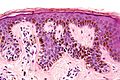
File:Dysplastic_nevus_-_add_-_high_mag.jpg|Dysplastic nevus under high magnification | |||
File:Dermatoscope1.JPG|Dermatoscope used for examining skin lesions | |||
File:Dermatoscope.jpg|Dermatoscope | |||
File:WB032021.JPG|Dysplastic nevus | |||
File:Pie_chart_of_incidence_and_malignancy_of_pigmented_skin_lesions.png|Pie chart of incidence and malignancy of pigmented skin lesions | |||
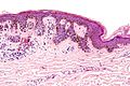
File:Dysplastic_nevus_-_low_mag.jpg|Dysplastic nevus under low magnification | |||
File:Dysplastic_nevus_-_intermed_mag.jpg|Dysplastic nevus under intermediate magnification | |||
File:Dysplastic_nevus_-_add_-_very_high_mag.jpg|Dysplastic nevus under very high magnification | |||
</gallery> | |||
Revision as of 11:37, 18 February 2025
Dysplastic nevus is a type of mole that is larger than normal (larger than a pencil eraser) and irregular in shape. It tends to have uneven color with dark brown centers and lighter, uneven edges. These moles tend to be hereditary (inherited from parents). People with dysplastic nevi may have more than 100 moles and have a greater chance of developing melanoma, a serious form of skin cancer.
Causes
The exact cause of dysplastic nevus is not known. However, it is believed to be influenced by a combination of genetic and environmental factors. Exposure to ultraviolet (UV) radiation from the sun and from tanning lamps and beds is a risk factor for all types of moles.
Symptoms
Dysplastic nevi often look different from common moles. A dysplastic nevus can have a mixture of several colors, from pink to dark brown. It is usually flat with a smooth, slightly scaly, or pebbly surface, and it has an irregular edge that may fade into the surrounding skin.
Diagnosis
A dermatologist can often identify a dysplastic nevus by its distinctive appearance. If there is any doubt, the dermatologist will perform a biopsy to make a definitive diagnosis.
Treatment
If a dysplastic nevus is suspected to be developing into a melanoma, it should be removed. However, if a dysplastic nevus does not change over time, there is no need for treatment.
Prevention
The best way to prevent dysplastic nevus and skin cancer is to protect the skin from the sun and other sources of UV radiation.
See also
References
<references />
|
|
 |
-
 Dysplastic nevus under high magnification
 -
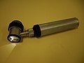 Dermatoscope used for examining skin lesions
 -
 Dermatoscope
 -
 Dysplastic nevus
 -
 Pie chart of incidence and malignancy of pigmented skin lesions
 -
 Dysplastic nevus under low magnification
 -
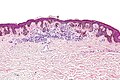 Dysplastic nevus under intermediate magnification
 -
 Dysplastic nevus under very high magnification


