Oesophagostomum: Difference between revisions
CSV import Tags: mobile edit mobile web edit |
CSV import |
||
| Line 22: | Line 22: | ||
{{medicine-stub}} | {{medicine-stub}} | ||
<gallery> | |||
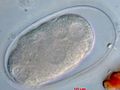
File:Oesophagostomum_larva_(micrograph).jpg|Oesophagostomum larva (micrograph) | |||
File:Parasite140080-fig3_Gastrointestinal_parasites_in_seven_primates_of_the_Taï_National_Park_-_Helminths_Figure_3e.jpg|Gastrointestinal parasites in seven primates of the Taï National Park - Helminths Figure 3e | |||
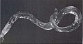
File:Oesophagostomum_Cylinder1.JPG|Oesophagostomum | |||
File:Oesophagostomum_life_cycle.jpg|Oesophagostomum life cycle | |||
</gallery> | |||
Latest revision as of 04:45, 18 February 2025
Oesophagostomum is a genus of parasitic nematodes, belonging to the family Strongylidae. These parasites are known to infect the intestines of various mammals, including humans, pigs, and ruminants, causing a disease known as oesophagostomiasis or nodular worm infection. The life cycle of Oesophagostomum species involves the development of larvae within the environment before infecting a new host, typically through ingestion of contaminated food or water.
Life Cycle[edit]
The life cycle of Oesophagostomum begins with the excretion of eggs in the feces of an infected host. These eggs hatch in the environment, releasing larvae that undergo several developmental stages. The infective larvae are then ingested by a new host. Once inside the host's gastrointestinal tract, the larvae penetrate the intestinal wall, forming nodules where they develop into adult worms. The adult worms then mate, and the females lay eggs, which are excreted in the host's feces, completing the cycle.
Pathogenesis and Clinical Signs[edit]
Infection with Oesophagostomum can lead to the formation of nodules in the intestinal wall, which can cause a variety of symptoms depending on the severity of the infection. In mild cases, infection may be asymptomatic. However, in more severe cases, symptoms can include diarrhea, abdominal pain, weight loss, and malnutrition. In ruminants, heavy infections can lead to reduced productivity and growth.
Diagnosis and Treatment[edit]
Diagnosis of oesophagostomiasis is typically made through the detection of eggs or larvae in fecal samples using microscopic examination. Treatment involves the administration of anthelmintic drugs, which are effective in killing the worms. Preventive measures include proper sanitation and hygiene practices to reduce environmental contamination with feces and the risk of infection.
Epidemiology[edit]
Oesophagostomum species are found worldwide, with a higher prevalence in tropical and subtropical regions where sanitation and hygiene practices may be inadequate. The distribution and prevalence of infection are influenced by factors such as climate, host species, and environmental contamination.
Prevention[edit]
Prevention of oesophagostomiasis involves controlling environmental contamination with feces to reduce the risk of infection. This can be achieved through the implementation of proper sanitation and hygiene practices, including the safe disposal of animal and human feces. Regular deworming of domestic animals can also help reduce the environmental burden of infective larvae.

-
 Oesophagostomum larva (micrograph)
 -
 Gastrointestinal parasites in seven primates of the Taï National Park - Helminths Figure 3e
 -
 Oesophagostomum
 -
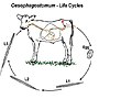 Oesophagostomum life cycle
.jpg)


