Spin echo: Difference between revisions
CSV import Tags: mobile edit mobile web edit |
CSV import |
||
| Line 30: | Line 30: | ||
{{Medicine-stub}} | {{Medicine-stub}} | ||
{{Physics-stub}} | {{Physics-stub}} | ||
<gallery> | |||
File:HahnEcho_GWM.gif|Spin_echo | |||
File:SpinEcho_GWM_stills.jpg|Spin_echo | |||
File:SpinEcho2_GWM.gif|Spin_echo | |||
File:GWM_HahnEchoDecay.gif|Spin_echo | |||
File:X-ray_of_subtle_compressive_hip_fracture,_labeled.jpg|X-ray of subtle compressive hip fracture, labeled | |||
File:CT_of_subtle_compressive_hip_fracture.jpg|CT of subtle compressive hip fracture | |||
File:T1_TSE_MRI_of_hip_fracture.jpg|T1 TSE MRI of hip fracture | |||
</gallery> | |||
Latest revision as of 11:24, 18 February 2025
Spin Echo is a fundamental concept in Magnetic Resonance Imaging (MRI) and Nuclear Magnetic Resonance (NMR) Spectroscopy, pivotal in the field of medical imaging and chemical analysis. It refers to a technique used to refocus magnetic resonance signals that have dephased due to inhomogeneities in the magnetic field, thereby enhancing signal intensity and improving image or spectrum quality.
Overview[edit]
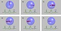
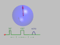
The spin echo technique was first introduced by Erwin Hahn in 1950. It involves the application of a series of radiofrequency (RF) pulses to a sample placed within a magnetic field. The initial pulse, typically a 90-degree pulse, flips the magnetization of the nuclei in the sample from its equilibrium position into the transverse plane. This is followed by a 180-degree pulse that inverts the spin system, causing the spins that were dephasing to rephase and produce an echo signal at a time TE (Echo Time) after the initial pulse.
Mechanism[edit]
The phenomenon of spin echo arises due to the interactions between the magnetic moments of the nuclei and the external magnetic field. After the initial RF pulse, the magnetic moments begin to precess around the magnetic field axis. Due to slight variations in the magnetic field across the sample (field inhomogeneities), the precessing spins start to dephase, leading to a decrease in the net transverse magnetization and, consequently, the signal. The 180-degree pulse effectively reverses the phase dispersion, causing the spins to rephase and generate a detectable echo signal.
Applications[edit]
Spin echo sequences are widely used in MRI for creating contrast between different tissues and detecting abnormalities. They are particularly useful for T2-weighted imaging, where the contrast is based on differences in the T2 relaxation times of tissues. This makes spin echo sequences valuable for diagnosing various medical conditions, including brain disorders, musculoskeletal injuries, and tumors.
In NMR spectroscopy, spin echo techniques help in measuring the relaxation times of nuclei, which can provide insights into the molecular dynamics and structure of the sample. They are also used to eliminate unwanted signals and artifacts, improving the clarity and resolution of the NMR spectrum.
Advantages and Limitations[edit]
The primary advantage of the spin echo technique is its ability to correct for field inhomogeneities, leading to clearer and more accurate images or spectra. However, spin echo sequences are generally longer than other sequences, such as gradient echo, making them more susceptible to motion artifacts. Additionally, the requirement for multiple RF pulses increases the specific absorption rate (SAR), which can be a concern in clinical MRI applications.
See Also[edit]
- Magnetic Resonance Imaging (MRI)
- Nuclear Magnetic Resonance (NMR) Spectroscopy
- T2 Relaxation Time
- Radiofrequency Pulse
- Magnetic Field Inhomogeneity
References[edit]
<references/>
This medical article is a stub. You can help Wikipedia by adding missing information. |
This physics-related article is a stub. You can help Wikipedia by adding missing information. |
-
 Spin_echo
 -
 Spin_echo
 -
 Spin_echo
 -
 Spin_echo
 -
 X-ray of subtle compressive hip fracture, labeled
 -
 CT of subtle compressive hip fracture
 -
 T1 TSE MRI of hip fracture


