Triangle of auscultation: Difference between revisions
CSV import Tags: mobile edit mobile web edit |
CSV import |
||
| (One intermediate revision by the same user not shown) | |||
| Line 1: | Line 1: | ||
{{Short description|Anatomical region on the back used for auscultation}} | |||
{{Use dmy dates|date=October 2023}} | |||
The triangle of auscultation is a | The '''triangle of auscultation''' is a small anatomical region on the back where the muscles are relatively thin, allowing for clearer auscultation of the lungs. This area is clinically significant as it provides a window for listening to respiratory sounds with a [[stethoscope]]. | ||
==Anatomy== | |||
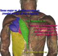
The triangle of auscultation is bordered by three muscles: | |||
* The [[latissimus dorsi]] forms the inferior border. | |||
* The [[trapezius]] forms the medial border. | |||
* The [[scapula]], specifically the medial border of the scapula, forms the lateral border. | |||
The floor of the triangle is formed by the [[rhomboid major]] muscle. When the scapula is protracted, the triangle becomes more pronounced, making it easier to auscultate the underlying lung tissue. | |||
==Clinical significance== | |||
The triangle of auscultation is used by healthcare professionals to listen to the sounds of the lungs. It is particularly useful for detecting [[breath sounds]] and identifying any abnormalities such as [[wheezing]], [[crackles]], or [[rales]]. The thin musculature in this area allows for clearer transmission of sound from the lungs to the stethoscope. | |||
==History== | |||
The triangle of auscultation was first described in the 19th century by anatomists who noted its clinical utility. It has since become a standard part of the physical examination of the respiratory system. | |||
==Images== | |||
[[File:VHM_Triangle_of_Auscultation.png|thumb|Diagram showing the location of the triangle of auscultation on the back.]] | |||
[[File:VHM_Triangle_of_Auscultation_CS.png|thumb|Cross-section illustrating the anatomical borders of the triangle of auscultation.]] | |||
==Related pages== | |||
* [[Auscultation]] | |||
* [[Respiratory system]] | |||
* [[Physical examination]] | |||
==References== | |||
{{Reflist}} | |||
[[Category:Anatomy]] | [[Category:Anatomy]] | ||
[[Category: | [[Category:Respiratory system]] | ||
<gallery> | |||
File:VHM_Triangle_of_Auscultation.png|Triangle of auscultation | |||
File:VHM_Triangle_of_Auscultation_CS.png|Triangle of auscultation | |||
</gallery> | |||
Latest revision as of 01:36, 18 February 2025
Anatomical region on the back used for auscultation
The triangle of auscultation is a small anatomical region on the back where the muscles are relatively thin, allowing for clearer auscultation of the lungs. This area is clinically significant as it provides a window for listening to respiratory sounds with a stethoscope.
Anatomy[edit]
The triangle of auscultation is bordered by three muscles:
- The latissimus dorsi forms the inferior border.
- The trapezius forms the medial border.
- The scapula, specifically the medial border of the scapula, forms the lateral border.
The floor of the triangle is formed by the rhomboid major muscle. When the scapula is protracted, the triangle becomes more pronounced, making it easier to auscultate the underlying lung tissue.
Clinical significance[edit]
The triangle of auscultation is used by healthcare professionals to listen to the sounds of the lungs. It is particularly useful for detecting breath sounds and identifying any abnormalities such as wheezing, crackles, or rales. The thin musculature in this area allows for clearer transmission of sound from the lungs to the stethoscope.
History[edit]
The triangle of auscultation was first described in the 19th century by anatomists who noted its clinical utility. It has since become a standard part of the physical examination of the respiratory system.
Images[edit]


Related pages[edit]
References[edit]
<references group="" responsive="1"></references>
-
 Triangle of auscultation
 -
 Triangle of auscultation
