Atrioventricular septum: Difference between revisions
CSV import |
CSV import Tags: mobile edit mobile web edit |
||
| Line 29: | Line 29: | ||
[[Category:Anatomy]] | [[Category:Anatomy]] | ||
{{anatomy-stub}} | {{anatomy-stub}} | ||
<gallery> | |||
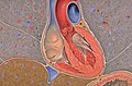
File:Gray_internal_structure_of_heart.png|Internal structure of the heart | |||
File:Heart_coronal_xs.jpg|Coronal section of the heart | |||
</gallery> | |||
Latest revision as of 05:00, 18 February 2025
Atrioventricular Septum
The atrioventricular septum is a vital component of the heart's anatomy, separating the atria and ventricles. It plays a crucial role in the heart's function, ensuring the proper flow of blood between the heart's chambers and preventing the mixing of oxygenated and deoxygenated blood.
Structure[edit]
The atrioventricular septum is composed of two parts: the membranous part and the muscular part. The membranous part is the smaller and thinner portion, located at the base of the septum. The muscular part, also known as the septum inferius, is the larger and thicker portion, extending from the base to the apex of the heart.
Function[edit]
The primary function of the atrioventricular septum is to separate the atria and ventricles, preventing the direct flow of blood between these chambers. This ensures that the blood follows the correct path through the heart, from the atria to the ventricles, and then out to the body or lungs.
Clinical Significance[edit]
Defects in the atrioventricular septum can lead to serious heart conditions. Atrioventricular septal defect (AVSD) is a congenital heart defect where there are holes in the atrioventricular septum and abnormalities in the atrioventricular valves. This can result in the mixing of oxygenated and deoxygenated blood, leading to symptoms such as shortness of breath, fatigue, and cyanosis.
See Also[edit]
References[edit]
<references group="" responsive="1"></references>

-
 Internal structure of the heart
 -
 Coronal section of the heart


