Basal plate (neural tube): Difference between revisions
CSV import Tags: mobile edit mobile web edit |
CSV import |
||
| Line 33: | Line 33: | ||
{{anatomy-stub}} | {{anatomy-stub}} | ||
== Basal plate (neural tube) gallery == | |||
<gallery> | |||
File:Gray646.png|Gray646 | |||
File:Human embryo 8 weeks 9.JPG|Human embryo 8 weeks 9 | |||
</gallery> | |||
Latest revision as of 05:02, 3 March 2025
Basal Plate (Neural Tube)
The Basal Plate is a region of the neural tube that gives rise to motor neurons and interneurons. It is located on the ventral side of the neural tube, opposite the alar plate, which is responsible for sensory neurons. The basal plate and alar plate are separated by the sulcus limitans.
Development[edit]
The basal plate forms during the neurulation process, when the neural plate folds in on itself to form the neural tube. The cells in the basal plate are specified to become motor neurons and interneurons due to the influence of sonic hedgehog (Shh), a morphogen secreted by the notochord and the floor plate.
Function[edit]
The basal plate gives rise to motor neurons, which control voluntary muscle movement, and interneurons, which relay signals between sensory and motor neurons. The motor neurons extend their axons out of the neural tube and into the developing muscles, while the interneurons remain within the central nervous system.
Clinical Significance[edit]
Abnormalities in the development of the basal plate can lead to a variety of neurological disorders, including spina bifida and anencephaly. These conditions are often caused by a failure of the neural tube to close properly during development.
See Also[edit]
References[edit]
<references />

Basal plate (neural tube) gallery[edit]
-
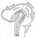 Gray646
 -
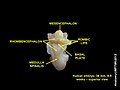 Human embryo 8 weeks 9


