Tzanck test: Difference between revisions
CSV import |
CSV import |
||
| (One intermediate revision by the same user not shown) | |||
| Line 1: | Line 1: | ||
{{Short description|A diagnostic test used in dermatology}} | |||
{{Use dmy dates|date=October 2023}} | |||
== | == Tzanck Test == | ||
The | The '''Tzanck test''' is a diagnostic tool used in [[dermatology]] to identify certain skin infections and conditions. It involves the microscopic examination of cells scraped from the base of a vesicular or bullous lesion. The test is named after the French dermatologist [[Arnault Tzanck]], who first described it. | ||
== | == Procedure == | ||
The Tzanck test is performed by scraping the base of a blister or vesicle with a scalpel blade. The collected material is then smeared onto a glass slide, air-dried, and stained with a [[Giemsa stain]] or [[Wright's stain]]. The stained slide is examined under a microscope for the presence of specific cellular changes. | |||
== | == Diagnostic Uses == | ||
The Tzanck test is primarily used to diagnose: | |||
* [[Herpes simplex virus]] (HSV) infections: The presence of multinucleated giant cells and acantholytic cells is indicative of HSV infections. | |||
* [[Varicella zoster virus]] (VZV) infections: Similar to HSV, VZV infections also show multinucleated giant cells. | |||
* [[Pemphigus vulgaris]]: Acantholytic cells are characteristic of this autoimmune blistering disorder. | |||
* [[Cutaneous leishmaniasis]]: The test can reveal intracellular and extracellular [[Leishmania]] parasites. | |||
== | == Limitations == | ||
While the Tzanck test can provide rapid results, it is not specific for any particular virus or condition. It cannot distinguish between HSV and VZV infections, and further testing, such as [[polymerase chain reaction]] (PCR) or viral culture, may be necessary for definitive diagnosis. | |||
== | == Related Pages == | ||
* [[Herpes simplex]] | |||
* [[Varicella zoster virus]] | |||
* [[Pemphigus]] | |||
* [[Leishmaniasis]] | |||
== | == Gallery == | ||
[[File:Tzanck_test.png|thumb|right|A Tzanck test being performed.]] | |||
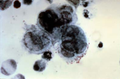
[[File:Acantholytic_cell_and_multinucleated_giant_cell_on_a_smear_taken_from_a_vesicular_lesion_of_herpes_simplex_infection.jpg|thumb|right|Acantholytic cell and multinucleated giant cell from a herpes simplex infection.]] | |||
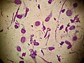
[[File:Extracellular_and_intracellular_leishmania_parasites_in_a_patient_with_cutaneous_leishmaniasis.jpg|thumb|right|Leishmania parasites in cutaneous leishmaniasis.]] | |||
[[File:Acantholytic_cells_on_a_smear_taken_from_a_patient_with_pemphigus_foliaceus.jpg|thumb|right|Acantholytic cells from pemphigus foliaceus.]] | |||
[[File:Tadpole_cells_on_a_smear_taken_from_a_patient_with_allergic_contact_dermatitis.jpg|thumb|right|Tadpole cells from allergic contact dermatitis.]] | |||
[[File:A_cluster_of_basaloid_cells_on_a_smear_taken_from_a_patient_with_basal_cell_carcinoma.jpg|thumb|right|Basaloid cells from basal cell carcinoma.]] | |||
== | == References == | ||
{{Reflist}} | |||
[[Category: | [[Category:Dermatology]] | ||
[[Category: | [[Category:Medical tests]] | ||
<gallery> | |||
File:Tzanck_test.png|Tzanck test | |||
File:Acantholytic_cell_and_multinucleated_giant_cell_on_a_smear_taken_from_a_vesicular_lesion_of_herpes_simplex_infection.jpg|Acantholytic cell and multinucleated giant cell on a smear taken from a vesicular lesion of herpes simplex infection | |||
File:Extracellular_and_intracellular_leishmania_parasites_in_a_patient_with_cutaneous_leishmaniasis.jpg|Extracellular and intracellular leishmania parasites in a patient with cutaneous leishmaniasis | |||
File:Acantholytic_cells_on_a_smear_taken_from_a_patient_with_pemphigus_foliaceus.jpg|Acantholytic cells on a smear taken from a patient with pemphigus foliaceus | |||
File:Tadpole_cells_on_a_smear_taken_from_a_patient_with_allergic_contact_dermatitis.jpg|Tadpole cells on a smear taken from a patient with allergic contact dermatitis | |||
File:A_cluster_of_basaloid_cells_on_a_smear_taken_from_a_patient_with_basal_cell_carcinoma.jpg|A cluster of basaloid cells on a smear taken from a patient with basal cell carcinoma | |||
</gallery> | |||
Latest revision as of 11:34, 18 February 2025
A diagnostic test used in dermatology
Tzanck Test[edit]
The Tzanck test is a diagnostic tool used in dermatology to identify certain skin infections and conditions. It involves the microscopic examination of cells scraped from the base of a vesicular or bullous lesion. The test is named after the French dermatologist Arnault Tzanck, who first described it.
Procedure[edit]
The Tzanck test is performed by scraping the base of a blister or vesicle with a scalpel blade. The collected material is then smeared onto a glass slide, air-dried, and stained with a Giemsa stain or Wright's stain. The stained slide is examined under a microscope for the presence of specific cellular changes.
Diagnostic Uses[edit]
The Tzanck test is primarily used to diagnose:
- Herpes simplex virus (HSV) infections: The presence of multinucleated giant cells and acantholytic cells is indicative of HSV infections.
- Varicella zoster virus (VZV) infections: Similar to HSV, VZV infections also show multinucleated giant cells.
- Pemphigus vulgaris: Acantholytic cells are characteristic of this autoimmune blistering disorder.
- Cutaneous leishmaniasis: The test can reveal intracellular and extracellular Leishmania parasites.
Limitations[edit]
While the Tzanck test can provide rapid results, it is not specific for any particular virus or condition. It cannot distinguish between HSV and VZV infections, and further testing, such as polymerase chain reaction (PCR) or viral culture, may be necessary for definitive diagnosis.
Related Pages[edit]
Gallery[edit]


References[edit]
<references group="" responsive="1"></references>
-
 Tzanck test
 -
 Acantholytic cell and multinucleated giant cell on a smear taken from a vesicular lesion of herpes simplex infection
 -
 Extracellular and intracellular leishmania parasites in a patient with cutaneous leishmaniasis
 -
 Acantholytic cells on a smear taken from a patient with pemphigus foliaceus
 -
 Tadpole cells on a smear taken from a patient with allergic contact dermatitis
 -
 A cluster of basaloid cells on a smear taken from a patient with basal cell carcinoma
