Ventilation/perfusion scan: Difference between revisions
CSV import Tag: Manual revert |
CSV import |
||
| (One intermediate revision by the same user not shown) | |||
| Line 1: | Line 1: | ||
''' | {{Short description|Medical imaging technique}} | ||
{{Use dmy dates|date=October 2023}} | |||
A '''ventilation/perfusion scan''' (V/Q scan) is a type of medical imaging using scintigraphy and medical isotopes to evaluate the circulation of air and blood within a patient's lungs. It is primarily used to help diagnose or rule out a [[pulmonary embolism]] (PE). | |||
==Procedure== | ==Procedure== | ||
The | The V/Q scan involves two parts: the ventilation scan and the perfusion scan. | ||
== | ===Ventilation scan=== | ||
In the ventilation scan, a patient inhales a radioactive gas or aerosol, such as xenon or technetium-labeled aerosol, which allows for imaging of the airways and distribution of air in the lungs. This part of the scan helps to assess the airflow within the lungs. | |||
== | ===Perfusion scan=== | ||
The perfusion scan involves the intravenous injection of a radioactive tracer, typically technetium-99m-labeled macroaggregated albumin. This tracer travels through the bloodstream and into the pulmonary circulation, allowing for imaging of blood flow in the lungs. Areas of the lung that receive less blood flow will appear as "cold spots" on the scan. | |||
== | ==Interpretation== | ||
The results of the V/Q scan are interpreted by comparing the ventilation and perfusion images. A mismatch between the two, where areas of the lung are ventilated but not perfused, may indicate a [[pulmonary embolism]]. | |||
== | ==Indications== | ||
The | The V/Q scan is most commonly used to diagnose or exclude pulmonary embolism, especially in patients who cannot undergo [[computed tomography pulmonary angiography]] (CTPA) due to allergies to contrast media or renal impairment. It may also be used to assess regional lung function in patients undergoing lung surgery. | ||
== | ==Limitations== | ||
While the V/Q scan is useful, it has limitations. It may not be as definitive as CTPA, and its accuracy can be affected by underlying lung conditions such as [[chronic obstructive pulmonary disease]] (COPD) or [[asthma]]. | |||
==Images== | |||
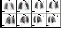
[[File:Ventperf.jpg|thumb|right|A typical ventilation/perfusion scan showing normal distribution of air and blood in the lungs.]] | |||
[[File:Pulmonary_embolism_scintigraphy_PLoS.png|thumb|right|A V/Q scan showing a mismatch indicative of a pulmonary embolism.]] | |||
==Related pages== | |||
* [[Pulmonary embolism]] | * [[Pulmonary embolism]] | ||
* [[Scintigraphy]] | |||
* [[Nuclear medicine]] | * [[Nuclear medicine]] | ||
[[Category:Medical | ==References== | ||
* "Ventilation/Perfusion Scan." RadiologyInfo.org. Accessed October 2023. | |||
* "Pulmonary Embolism: Diagnosis and Management." American Family Physician, 2023. | |||
[[Category:Medical imaging]] | |||
[[Category:Nuclear medicine]] | [[Category:Nuclear medicine]] | ||
<gallery> | |||
File:Ventperf.jpg|Ventilation/perfusion scan | |||
File:Pulmonary_embolism_scintigraphy_PLoS.png|Pulmonary embolism scintigraphy | |||
</gallery> | |||
Latest revision as of 00:46, 18 February 2025
Medical imaging technique
A ventilation/perfusion scan (V/Q scan) is a type of medical imaging using scintigraphy and medical isotopes to evaluate the circulation of air and blood within a patient's lungs. It is primarily used to help diagnose or rule out a pulmonary embolism (PE).
Procedure[edit]
The V/Q scan involves two parts: the ventilation scan and the perfusion scan.
Ventilation scan[edit]
In the ventilation scan, a patient inhales a radioactive gas or aerosol, such as xenon or technetium-labeled aerosol, which allows for imaging of the airways and distribution of air in the lungs. This part of the scan helps to assess the airflow within the lungs.
Perfusion scan[edit]
The perfusion scan involves the intravenous injection of a radioactive tracer, typically technetium-99m-labeled macroaggregated albumin. This tracer travels through the bloodstream and into the pulmonary circulation, allowing for imaging of blood flow in the lungs. Areas of the lung that receive less blood flow will appear as "cold spots" on the scan.
Interpretation[edit]
The results of the V/Q scan are interpreted by comparing the ventilation and perfusion images. A mismatch between the two, where areas of the lung are ventilated but not perfused, may indicate a pulmonary embolism.
Indications[edit]
The V/Q scan is most commonly used to diagnose or exclude pulmonary embolism, especially in patients who cannot undergo computed tomography pulmonary angiography (CTPA) due to allergies to contrast media or renal impairment. It may also be used to assess regional lung function in patients undergoing lung surgery.
Limitations[edit]
While the V/Q scan is useful, it has limitations. It may not be as definitive as CTPA, and its accuracy can be affected by underlying lung conditions such as chronic obstructive pulmonary disease (COPD) or asthma.
Images[edit]


Related pages[edit]
References[edit]
- "Ventilation/Perfusion Scan." RadiologyInfo.org. Accessed October 2023.
- "Pulmonary Embolism: Diagnosis and Management." American Family Physician, 2023.
-
 Ventilation/perfusion scan
 -
 Pulmonary embolism scintigraphy
