File:Steroidogenesis.svg

Original file (SVG file, nominally 1,245 × 1,105 pixels, file size: 224 KB)
Summary
| Description |
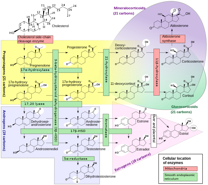
English: Enzymes, their cellular location, substrates and products in human steroidogenesis.
Shown also is the major classes of steroid hormones: progestagens, mineralocorticoids, glucocorticoids, androgens and estrogens. However, they partly overlap, e.g. mineralocorticoids and glucocorticoids. White circles indicate changes in molecular structure compared with precursors. For more information on interpretation of molecular structures, see structural formula. HSD: Hydroxysteroid dehydrogenase References:
Further reading:
See also: Polski: Enzymy biorące udział w steroidogenezie.
Uwagi: W miejscu gdzie stereochemia nie jest wyświetlana, jest taka sama jak dla Cholesterol lub w pokazanej powyżej grupie. HSD: Dehydrogenaza hydroksysteroiduРусский: Ферменты, участвующие в стероидогенезе человека, с указанием субстратов и продуктов, а также локализации их активности. Происходящие изменения в структуре молекул отмечены белыми кружочками. По сборнику "Медицинская физиология" (Walter F. Boron, Emile L. Boulpaep, 2003 год).
日本語: この図はヒトのステロイドホルモン生成における酵素と酵素が存在する細胞器官、基質ならびに生成物の全体図である。またこの図は(コレステロールから)プロゲステロン、鉱質コルチコイド、糖質コルチコイド、そしてアンドロゲンおよびエストロゲンに至る主要なステロイドを記述している。
図に示すとおりそれぞれ(生合成の過程で)オーバーラップするものである。 図中、白い円で囲んだところは基質と比較した分子構造の変化した箇所を示す。 なお、HSD は、ヒドロキシステロイドデヒドロゲナーゼを表す。 |
| Date | |
| Source | Häggström M, Richfield D (2014). "Diagram of the pathways of human steroidogenesis". WikiJournal of Medicine 1 (1). DOI:10.15347/wjm/2014.005. ISSN 20024436. |
| Author |
David Richfield (Slashme) and Mikael Häggström. Derived from previous version by Hoffmeier and Settersr. In external use, this diagram may be cited as:
|
| Other versions |
 Raster (.png) version is available Translations
|
| SVG development | This structural formula was created with Inkscape. |
.svg)
.svg)


|
EvolutionA short flashback of some milestones the evolution of the picture, demonstrating the power of free licensing where anybody can contribute to making things better.  .svg) .gif)  |
Licensing
| This file is made available under the Creative Commons CC0 1.0 Universal Public Domain Dedication. | |
| The person who associated a work with this deed has dedicated the work to the public domain by waiving all of their rights to the work worldwide under copyright law, including all related and neighboring rights, to the extent allowed by law. You can copy, modify, distribute and perform the work, even for commercial purposes, all without asking permission.
|
File history
Click on a date/time to view the file as it appeared at that time.
| Date/Time | Thumbnail | Dimensions | User | Comment | |
|---|---|---|---|---|---|
| current | 10:52, 28 October 2022 | | 1,245 × 1,105 (224 KB) | wikimediacommons>Uxue García | File uploaded using svgtranslate tool (https://svgtranslate.toolforge.org/). Added translation for eu. |
File usage
There are no pages that use this file.