File:Human brain left dissected midsagittal view description 2.JPG
Jump to navigation
Jump to search


No higher resolution available.
Human_brain_left_dissected_midsagittal_view_description_2.JPG (701 × 488 pixels, file size: 50 KB, MIME type: image/jpeg)
Summary
| Description |
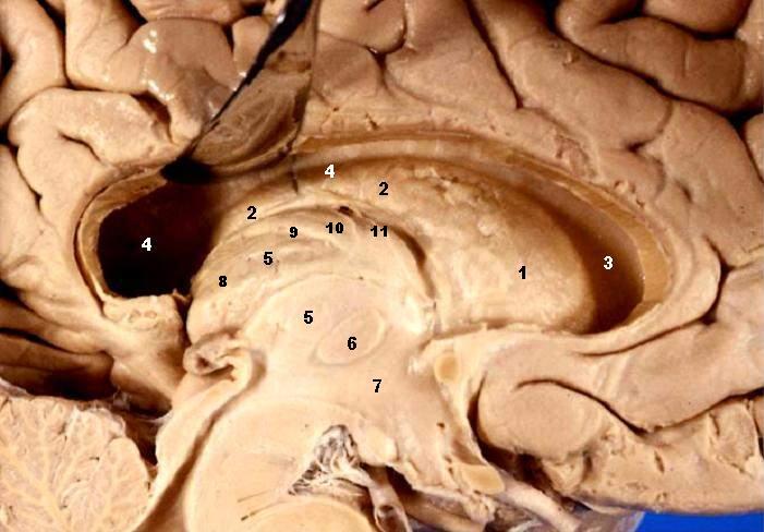
Human brain left dissected - midsagittal view Fornix & Septum Pellucidum Resected – Lateral Ventricle Exposed The head and body of the Caudate nucleus produce a large elevation in the lateral wall of the anterior horn and body of the lateral ventricle
|
| Date | |
| Source | http://www.healcentral.org/healapp/showMetadata?metadataId=40566 (Internet Archive of file description page) |
| Author |
John A Beal, PhD Dep't. of Cellular Biology & Anatomy, Louisiana State University Health Sciences Center Shreveport |
| Permission (Reusing this file) |
CC-BY |
| Other versions |
 |
Licensing
This file is licensed under the Creative Commons Attribution 2.5 Generic license.
- You are free:
- to share – to copy, distribute and transmit the work
- to remix – to adapt the work
- Under the following conditions:
- attribution – You must give appropriate credit, provide a link to the license, and indicate if changes were made. You may do so in any reasonable manner, but not in any way that suggests the licensor endorses you or your use.
This file, which was originally posted to
http://www.healcentral.org/healapp/showMetadata?metadataId=40566, was reviewed on 25 September 2013 by reviewer Eleassar, who confirmed that it was available there under the stated license on that date.
|
| Annotations | This image is annotated: View the annotations at Commons |
File history
Click on a date/time to view the file as it appeared at that time.
| Date/Time | Thumbnail | Dimensions | User | Comment | |
|---|---|---|---|---|---|
| current | 22:14, 22 June 2006 | | 701 × 488 (50 KB) | wikimediacommons>Patho | {{Information| |Description='''Human brain left dissected - midsagittal view''' Fornix & Septum Pellucidum Resected – Lateral Ventricle Exposed The head and body of the Caudate nucleus produce a large elevation in the lateral wall of the anterior hor |
File usage
There are no pages that use this file.