File:Histopathology of a pheochromocytoma with coagulative necrosis, with immunostaining.jpg
Jump to navigation
Jump to search


Size of this preview: 800 × 571 pixels. Other resolutions: 320 × 228 pixels | 640 × 457 pixels | 1,024 × 731 pixels | 1,232 × 879 pixels.
Original file (1,232 × 879 pixels, file size: 473 KB, MIME type: image/jpeg)
Summary
| Description |
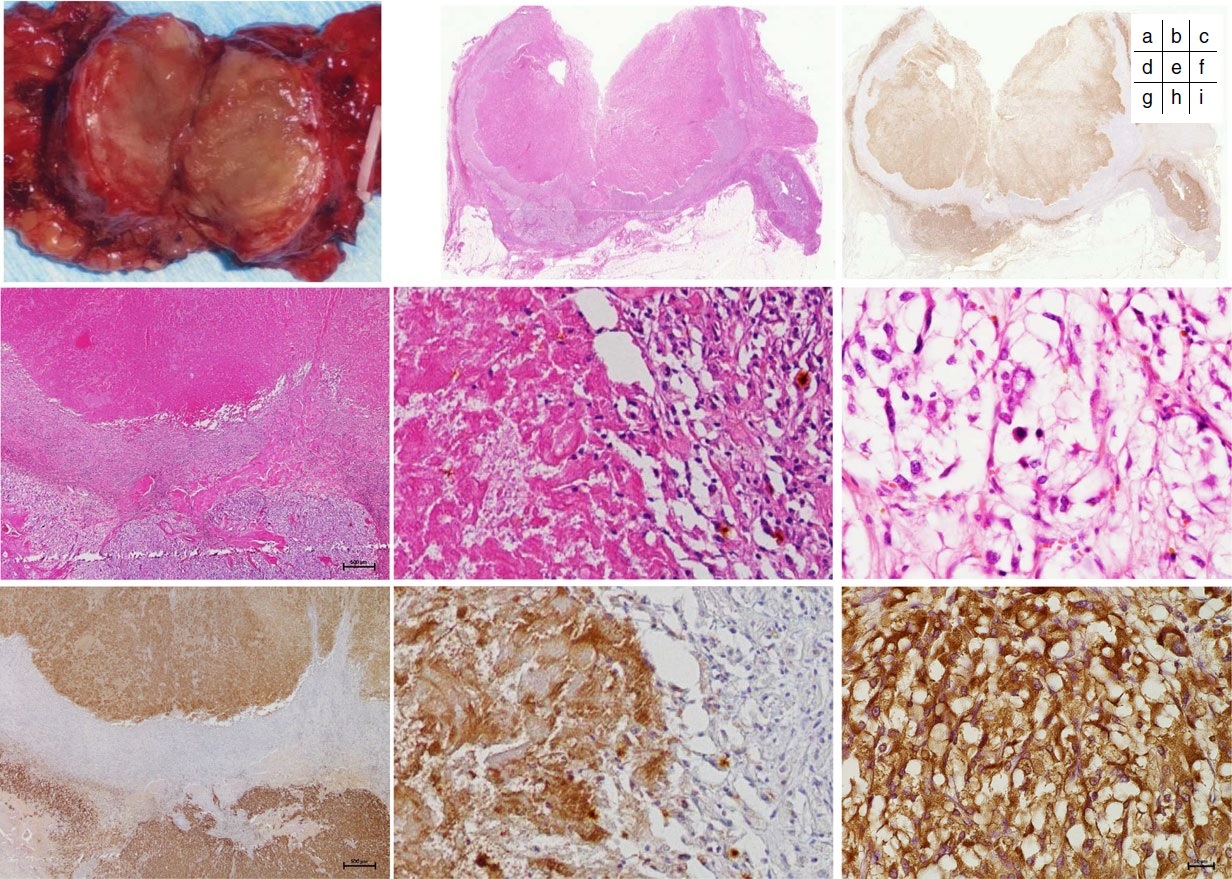
English: Original caption: Histopathological findings of the resected left adrenal gland (September 2009). a Gross appearance of the cut surface of the left adrenal tumor 3 cm in size showed the inferior surface to be necrotic. b−i Microscopic examination of the left adrenal tumor (b, d−f; hematoxylin and eosin staining. c, g−i; chromogranin A staining). Nontumoral adrenal gland in the right lower corner, and well-encapsulated tumor in the remainder of the photograph (b). The tumor had a large area of coagulative necrosis in the center. The necrotic material contained morphologically ghost cells (d, e) and was immunohistochemically markedly positive for chromogranin A (c, g, h). There were numerous hemosiderin-laden macrophages and histiocytes accompanied by vascular proliferation in the region adjacent to the area of necrosis (e, h). The viable region along the periphery of the tumor contained numerous cells undergoing pyknosis (f), and the cytoplasm of the tumor cells was positive for chromogranin A staining (i) |
| Date | |
| Source |
(2016). "Histopathological analysis of spontaneous large necrosis of adrenal pheochromocytoma manifested as acute attacks of alternating hypertension and hypotension: a case report". Journal of Medical Case Reports 10 (1). DOI:10.1186/s13256-016-1068-3. ISSN 1752-1947. - "This article is distributed under the terms of the Creative Commons Attribution 4.0 International License (http://creativecommons.org/licenses/by/4.0/)," |
| Author | Nobumasa Ohara, Yasuyuki Uemura, Naomi Mezaki, Keita Kimura, Masanori Kaneko, Hirohiko Kuwano, Katsuya Ebe, Toshio Fujita, Takeshi Komeyama, Hiroyuki Usuda, Yuto Yamazaki, Takashi Maekawa, Hironobu Sasano, Kenzo Kaneko & Kyuzi Kamoi |
| Other versions |
 |
Licensing
This file is licensed under the Creative Commons Attribution 4.0 International license.
- You are free:
- to share – to copy, distribute and transmit the work
- to remix – to adapt the work
- Under the following conditions:
- attribution – You must give appropriate credit, provide a link to the license, and indicate if changes were made. You may do so in any reasonable manner, but not in any way that suggests the licensor endorses you or your use.
File history
Click on a date/time to view the file as it appeared at that time.
| Date/Time | Thumbnail | Dimensions | User | Comment | |
|---|---|---|---|---|---|
| current | 13:28, 3 January 2020 | | 1,232 × 879 (473 KB) | wikimediacommons>Mikael Häggström | User created page with UploadWizard |
File usage
There are no pages that use this file.